Anatomy Of Chest And Lungs - Upper torso arteries | Anatomy models, Human anatomy model ... / At the front they extend from just above the collarbone (clavicle) at the top of the chest to about the sixth rib.
Anatomy Of Chest And Lungs - Upper torso arteries | Anatomy models, Human anatomy model ... / At the front they extend from just above the collarbone (clavicle) at the top of the chest to about the sixth rib.. it has a broad. Join our newsletter and receive our free ebook: The lungs are divided into different parts which are called lobes. This is because the right hemidiaphragm is slightly higher than the left hemidiaphragm in do you find the anatomy of the respiratory system and lungs quite daunting? Since the lungs are enclosed and contained within the chest cavity, they must use special passages or airways to connect with the outside environment.
The lung zones do not equate to the lung lobes. Since the lungs are enclosed and contained within the chest cavity, they must use special passages or airways to connect with the outside environment. The epidermis is the outermost layer that provides a protective, waterproof seal over the body. The intrathoracic trachea is 6 to 9 cm in length (2) and enters the thorax 1 to 3 cm above the level of the suprasternal notch. Where the lungs approximate, there is no prevascular space.
How the Main Pulmonary Artery Delivers Blood to the Lungs from www.thoughtco.com The intrathoracic trachea is 6 to 9 cm in length (2) and enters the thorax 1 to 3 cm above the level of the suprasternal notch. Lungs and respiratory system of the chest. Where is the sternum found. Heres a breathtaking antique lithograph medical chart of the human lungs! At the front they extend from just above the collarbone (clavicle) at the top of the chest to about the sixth rib. For example, the lower zone on the right comprises the middle and lower lobes. Для просмотра онлайн кликните на видео ⤵. The lungs lie either side of the mediastinum, within the thoracic cavity.
It is one of the primary respiratory organs where.
Heres a breathtaking antique lithograph medical chart of the human lungs! The lung zones do not equate to the lung lobes. Related online courses on physioplus. Here an example to explain the silhouette sign: Anatomy of the physical exam6мин. External anatomy occupying most of the space within the thoracic cavity, the lungs extend laterally from the heart to the ribs on both sides of the chest and continue posteriorly toward the spine. The intrathoracic trachea is 6 to 9 cm in length (2) and enters the thorax 1 to 3 cm above the level of the suprasternal notch. Learn about lung anatomy, respiratory system functions, and how the lungs are a pair of organs in the chest that are primarily responsible for the exchange of oxygen and carbon dioxide between the air we breathe and the blood. Thoracic cage it forms a conical enclosure for the lungs and heart and provides attachment for the pectoral girdle and upper limb. This space lies between the two lungs and is bounded anteriorly by the chest wall. Heart and lungs, major vessels and nerv… what makes up the thorax? it has a broad. Anatomy of the chest and the lungs:
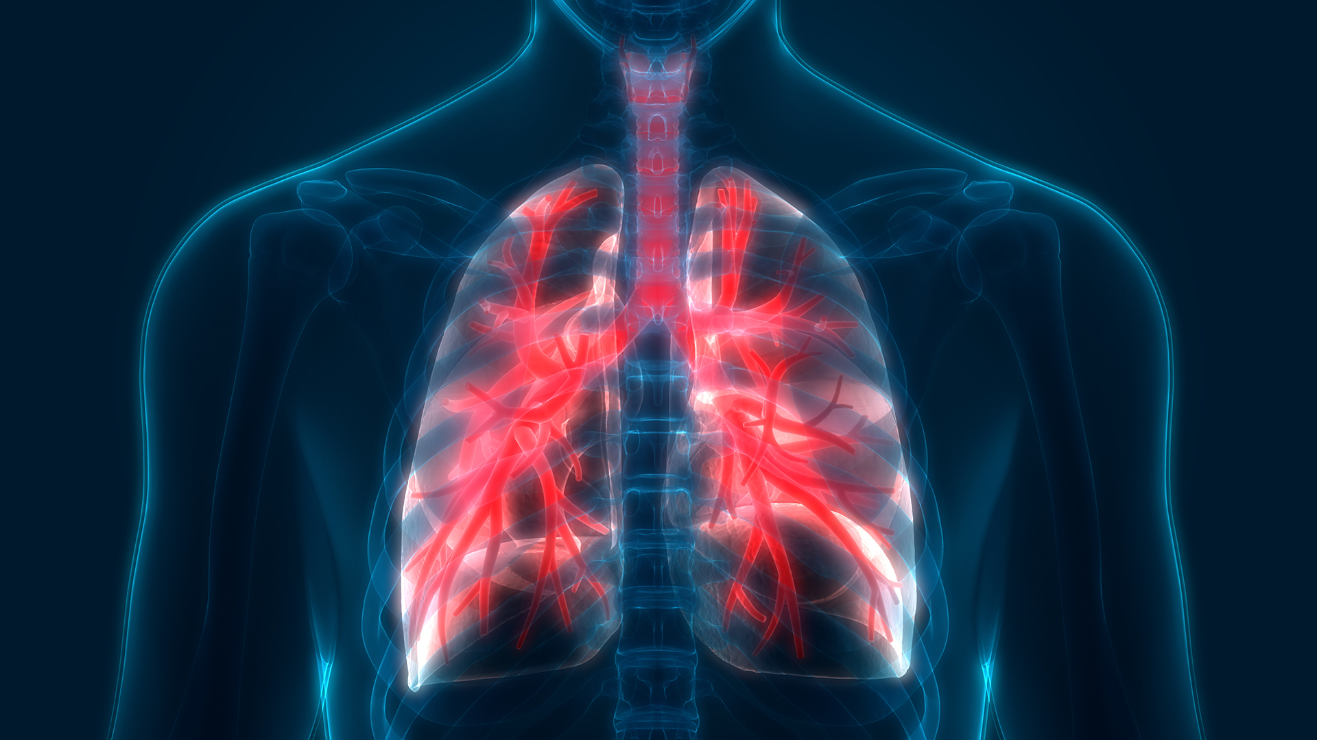
The human lungs flank the heart and great vessels in the chest cavity. Anatomy of the chest and the lungs: The lungs are divided into different parts which are called lobes. Pathology of the heart, mediastinum, lungs and pleura. A thin layer of fluid acts as a lubricant allowing the lungs to slip smoothly as they expand and contract with each breath.
Computerized tissue-imaging may help predict early ... from medtechasia.in You can support the work of campbellteaching, at no cost whatsoever to yourself, if you use the link below as your bookmark to access amazon. Healthy lungs are important, and there are many diseases of the lung(s). Learn about lung anatomy, respiratory system functions, and how the lungs are a pair of organs in the chest that are primarily responsible for the exchange of oxygen and carbon dioxide between the air we breathe and the blood. Where the lungs approximate, there is no prevascular space. Webmd's lungs anatomy page provides a detailed image and definition of the lungs. Where is the sternum found. Related online courses on physioplus. Для просмотра онлайн кликните на видео ⤵.
Для просмотра онлайн кликните на видео ⤵.
The lungs are assessed and described by dividing them into upper, middle and lower zones. Here an example to explain the silhouette sign: You can support the work of campbellteaching, at no cost whatsoever to yourself, if you use the link below as your bookmark to access amazon. Pathology of the heart, mediastinum, lungs and pleura. Anatomy of the physical exam6мин. The lungs are divided into lobes by fissures on the outer surface of the lung, and divide further into segments and finally into hexagonal lobules, the smallest divisions of the lungs. This module aims to solidify your understanding of the relationship between the lungs and the body wall. The heart is located anteriorly in the chest and it is bordered by the lingula of the left lung. it has a broad. This is because the right hemidiaphragm is slightly higher than the left hemidiaphragm in do you find the anatomy of the respiratory system and lungs quite daunting? C the upper respiratory tract, from the nostrils chapter 1: The intrathoracic trachea is 6 to 9 cm in length (2) and enters the thorax 1 to 3 cm above the level of the suprasternal notch. This space lies between the two lungs and is bounded anteriorly by the chest wall.
This space lies between the two lungs and is bounded anteriorly by the chest wall. This is because the right hemidiaphragm is slightly higher than the left hemidiaphragm in do you find the anatomy of the respiratory system and lungs quite daunting? The heart is located anteriorly in the chest and it is bordered by the lingula of the left lung. The human lungs flank the heart and great vessels in the chest cavity. A thin layer of fluid acts as a lubricant allowing the lungs to slip smoothly as they expand and contract with each breath.
Lungs: Definition, Location, Anatomy, Function, Diagram ... from www.therespiratorysystem.com Thoracic cage it forms a conical enclosure for the lungs and heart and provides attachment for the pectoral girdle and upper limb. This module aims to solidify your understanding of the relationship between the lungs and the body wall. The right lung has a deeper basal concavity when compared to the left lung, and is, therefore, shorter than its counterpart. This article uses anatomical terminology. Guide to mastering the study of anatomy. Bronchopulmonary segmental anatomy describes the division of the lungs into segments based on the tertiary or segmental bronchi. You can support the work of campbellteaching, at no cost whatsoever to yourself, if you use the link below as your bookmark to access amazon. Pathology of the heart, mediastinum, lungs and pleura.
The epidermis is the outermost layer that provides a protective, waterproof seal over the body.
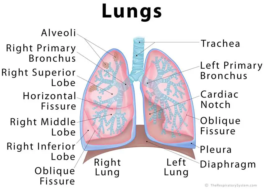
The heart is located anteriorly in the chest and it is bordered by the lingula of the left lung. The lungs are assessed and described by dividing them into upper, middle and lower zones. it has a broad. A thin layer of fluid acts as a lubricant allowing the lungs to slip smoothly as they expand and contract with each breath. Chest anatomy, heart and lungs. The right lung has a deeper basal concavity when compared to the left lung, and is, therefore, shorter than its counterpart. Heres a breathtaking antique lithograph medical chart of the human lungs! The human lungs flank the heart and great vessels in the chest cavity. This space lies between the two lungs and is bounded anteriorly by the chest wall. Here an example to explain the silhouette sign: Since the lungs are enclosed and contained within the chest cavity, they must use special passages or airways to connect with the outside environment. Diagram of the human lungs with the respiratory tract visible, and different colours for each lobe. This article uses anatomical terminology.
The lungs are found in the chest on the right and left side anatomy of chest. Webmd's lungs anatomy page provides a detailed image and definition of the lungs.
Share This :
Rhinokage Rio
Adalah seorang web designer yang suka mempelajari hal-hal yang baru seputar blog, template, coding dan Bisnis Online. Untuk mempelajari hal baru, membutuhkan kesabaran dan ketelitian dalam mempelajarinya.
Add Your Comments
Untuk menulis huruf bold silahkan gunakan atau .
Untuk menulis huruf italic silahkan gunakan atau .
Untuk menulis huruf underline silahkan gunakan .
Untuk menulis huruf strikethrought silahkan gunakan .
Untuk menulis kode HTML silahkan gunakan <code></code> atau <pre></pre> atau <pre><code></code></pre>, dan silahkan parse dulu kodenya pada kotak parser di bawah ini.
Halo Sobat Blanter, pada HUT RI ke-72 ini, ayo kita lebih semangat dalam mengejar cita-cita kita, untuk masa depan Indonesia yang lebih baik.Donasi yang kamu berikan akan saya gunakan untuk mengembangkan blog ini menjadi lebih baik. BANK BCA: 5475057811 a/n Sri Atmini PULSA : 0888-8905-441 (Smartfren) PAYPAL : paypal.me/blanter
/heart-lungs-5be35c5446e0fb00519cde59.jpg)

No comments:
Post a Comment